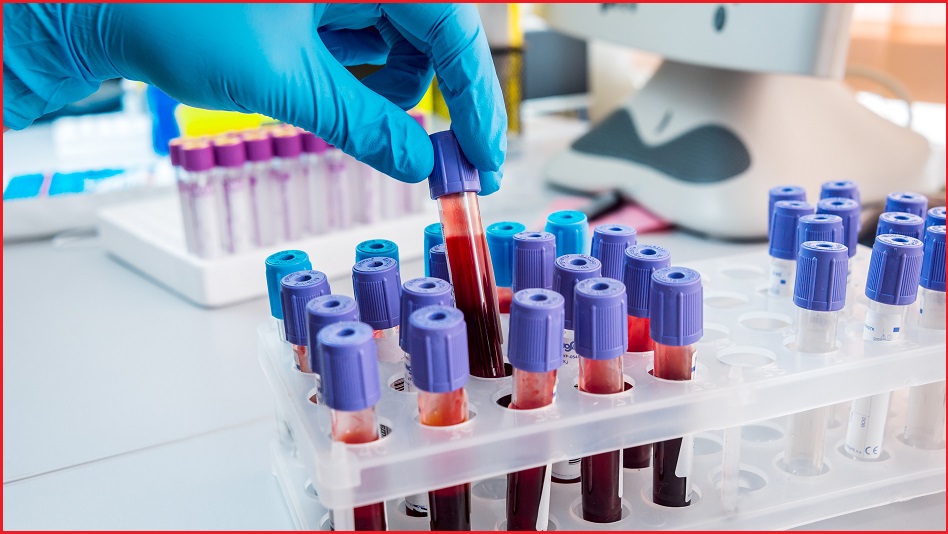

The processing of blood samples is set to become more efficient and accurate, after a Queensland pathology laboratory implemented an AI-powered microscope that uses pattern matching and image recognition to detect disease faster than human pathologists can.
In development for much of the past decade, the new machine – created in conjunction with the University of Queensland’s AI Collaboratory (AIC) and claimed to be a world first – automatically scans blood samples sent to the Brisbane central laboratory of pathology giant Sullivan Nicolaides Pathology (SNP), whose team of specialist pathologists have historically processed around 35,000 blood samples per day.
The microscope takes high-resolution images of each prepared slide, then uses AI algorithms to compare the features in the image to known images with which it has been trained by a human pathologist.
By analysing the shape of blood cells, the presence of other cells in the samples, and other telltale signs of disease, the machine can quickly flag samples as suspicious and send them for corroboration by human pathologists, along with automatically optimised photographs.
Use of the system has shown that the lab can look at ten times as many cells to classify a blood sample within the same amount of time, UQ AI Collaboratory professor Brian Lovell said, trumpeting its improved accuracy as the system was recently demonstrated.
“We used to look at 100 cells to classify [disease],” he explained. “Now we can look at 1000 cells, and you get a much better outcome.”
“The field of microscopy hasn’t changed much since germs were first identified in the 1800s,” he continued. “So what we’re doing is taking microscopy out of that 19th century paradigm into a 21st century paradigm.”
An objective pair of eyes
For all its automation benefits, the system still maintains a high degree of human oversight: human pathologists review flagged samples and continuously review and refine the system’s training set.
The AI microscope is part of a new breed of image-based pathology tools that – in the same way that AI is revolutionising applications like IVF embryo selection – is expected to rapidly take over much of the routine work at the world’s pathology laboratories.
Australia’s 24,000 pathology employees analyse around 500 million tests per year – but with a worldwide shortage of pathologists and a training path that takes 13 years to complete, skills pipeline issues remain a significant challenge even as the population, and therefore demand, grow.
Little wonder that a panel of pathology experts agreed AI will be “routinely and impactfully used” within the sector by 2030, a recent academic study found, with AI tools set to increase diagnostic accuracy, increase detection of rare events such as small metastases and small tumour foci, enable more quantitative analysis and improve the completeness, complexity, and quality of pathology reports.
Because its analysis is completely objective, the experts agreed, it is also valuable for standardising the diagnosis and grading of tumours – a process that currently suffers from high “interobserver variability”, meaning that different pathologists reach different conclusions when looking at the same sample.
“As pathology data get more complex, the discovery of meaningful patterns and relevant biomarkers for specific disease subtypes becomes more and more challenging,” one recent analysis noted, lauding AI’s ability to objectively complete “repetitive, subjective, or quantitative routine tasks” such as the “repetitive and error-prone” counting of hundreds of small cells in a sample.
“It’s like having a second pair of eyes, or a good trainee pathologist by your side,” noted Associate Professor Ewan Millar, a senior staff specialist histopathologist with NSW Health Pathology who has been investigating AI’s use in diagnosing prostate, breast and other cancers.
“Pathologists can look at hundreds of slides a day [and] finding cancerous tissue can be time-consuming,” Millar recently explained, noting that a human pathologist might often examine up to 75 slides to detect a single prostate cancer tumour of just 1mm in diameter.
“Some work can be like looking for a needle in a haystack,” he said, “but the AI can be trained to recognise pixel patterns [and] is capable of reviewing the slides within minutes.”
Another AIC project is already using AI to identify and classify skin disease such as melanoma.
“People require experience and training to analyse images,” explained Dr Shakes Chandra, a senior lecturer with the University of Queensland School of Electrical Engineering and Computer Science.
”AI speeds this up and allows us to process the data faster. Then, we can not only process the data but train the data to enable us to do more than we ever thought possible.”