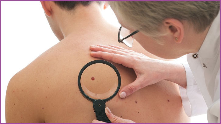

Machines for detecting skin cancer are being trialed in Australia next year in a telehealth pilot program that will help inform future melanoma-spotting algorithms.
Run out of the University of Queensland’s (UQ) Diamantina Institute, the program will see 15 walk-in skin cancer imaging machines installed in Victoria, New South Wales, and Queensland.
Professor Monika Janda from UQ said the research seeks to improve early the identification rates for Australia’s most common cancer.
“First and primarily this tool will provide clinicians with photographs for their own clinical diagnoses – it is invaluable for them to have total body imaging available for future reference,” she told Information Age.
The machines look similar to full-body airport scanners.
Once stripped off and standing inside the machine, patients are simultaneously photographed by 92 cameras.
After software renders and combines the images, the result is a digital avatar of the patient’s skin.
Crucially, this image can be examined remotely by expert dermatologists who, like many medical specialists, are few and far between in regional Australia.
Telehealth powered by this sort of imaging machine could be a gamechanger for identifying skin cancers – of which Australia has the one of the highest rates in the world.
The Australian Cancer Research Foundation (ACRF) provided a $9.9 million grant toward the program and is establishing the ACRF Australian Centre of Excellence in Melanoma Imaging and Diagnosis at UQ’s Diamantina Institute.
Kerry Strydom, CEO of the ACRF, said technology is a cornerstone of helping reduce the number of people who die by skin cancer.
“Cancer impacts every one of us,” she said.
“Through technology if we can find better ways to prevent, detect, and treat cancer, then we will be able to save lives.
“This is a very worthy recipient of a major grant and it will help diagnosis – and treatment – to come sooner for many Australians.”
Aside from testing the interopability and data privacy of these complex devices, the researchers will also look at augmenting them with image recognition algorithms.
Large amounts of de-identified data gathered from the first stage of research will help teach and improve those machine-learning algorithms and will build an Australian dataset that better takes into account the high levels of sun exposure and radiation damage our skin tends to have.
Already the International Skin Imaging Collaboration (ISIC) runs a yearly classification challenge which this year published open data of 33,000 images to train machine vision algorithms.
Ongoing research into machine learning for cancer detection has proved fruitful with a 2018 German research paper finding that a convolutional neural network outperformed some expert dermatologists at classifying skin cancers.
But while she is optimistic about the power of AI for cancer detection, Professor Janda thinks it is important to remember the human role in medicine.
“People have on average between 55 to upwards of 400 pigmented skin lesions – and even more non-pigmented lesions,” she said.
“In the future an AI could point out which lesions look okay and which might be harmful. It could also be a tool that clinicians use to check against when they are unsure.
“That could obviously save time and simplify things for clinicians – but AI will never replace them.
“We will always need a clinician to double-check things and be responsible for making final decisions.
"They are also the ones who have to be there talking with patients, explaining things, and making treatment plans.”