
An interactive virtual reality (VR) system has been created for patients undertaking MRI scans to make it easier for those who find them challenging, such as children, people with cognitive difficulties and claustrophobes.
And it all started with an unexpected gift.
After receiving VR goggles as a Christmas present, a researcher from the Centre for the Developing Brain at the King’s College London discovered first-hand how the strong immersive experience could also address difficulties around having an MRI scan.
This realisation that when someone is immersed in a VR environment, they are entirely unaware of their surroundings, led to the quest to create a system compatible with the MRI environment.
“We went on to develop the required new technology through a multi-disciplinary collaboration of colleagues with a background in computer science, engineering, optical science, clinical and physics,” Dr Tomoki Arichi, clinical senior lecturer at the Centre for the Developing Brain, King’s College London, said.
Why VR makes a difference
MRIs are an important element in clinical practice and medical research; however, patients undergoing these scans often experience anxiety and sometimes distress prior to and during scanning.
What makes this VR application unique is this it provides a full-body immersive experience.
“When people are using it, they are unaware of their surroundings and completely distracted from all the alien sensations associated with the scanning process,” Arichi explained to Information Age.
Typical VR setups are head-mounted using goggles or other form of headset along with head motion tracking technology that can create and maintain a visceral sense of a virtual world.
In these applications, VR encourages physical motion but it is physically incompatible for MRI scans where the patient lays on a narrow bed and is then enclosed in a solid tube-like structure inhibiting movement.
This new system uses gaze tracking, adapted from VR features used for camera view control and aiming, which monitors and interacts with a virtual world through MRI-compatible cameras.
To adapt it, the researchers developed a dedicated VR framework that includes a rich virtual world and gaze-controlled content.
In addition, by excluding the peripheral vision cues which anchor the person to their physical situation, it allows the VR experience to more completely replace the sensation of being inside an MRI scanner.
While the scan is being carried out, a video link to the carer or practitioner also projects a supportive presence into the virtual world.
They also hope that giving the patient control and providing ease of communication with a carer outside the scanner could potentially transform the MR examination, taking it “from a passive, boring and isolating experience into an active, engaging and interactive one”.
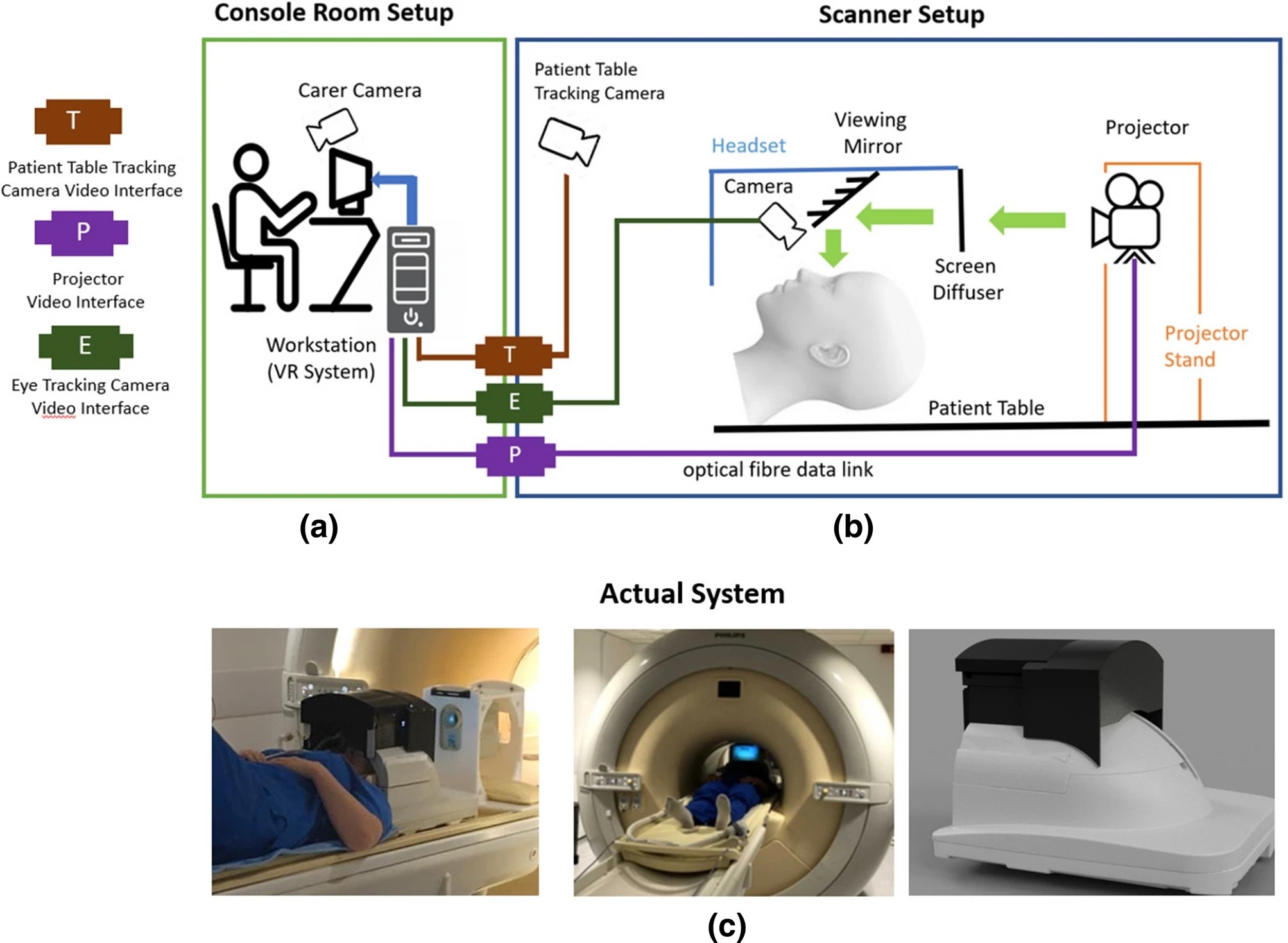
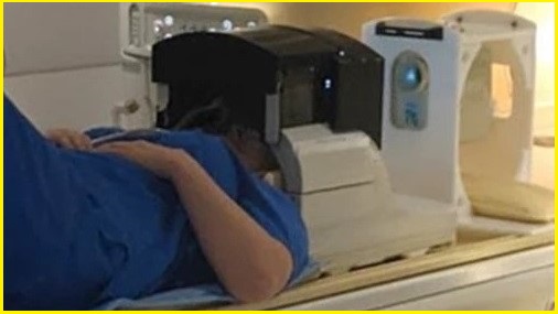
Top: Illustration of setup in the scanner and console rooms; bottom: system and scanner headset. Image: Centre for the Developing Brain
Tailored content is key
The researchers believe that replacing the images seen by the patient or research with tailored VR content that is immersive and interactive could achieve a radically different subjective experience.
When a person starts using the system, they first enter an initial introduction area and then a lobby to navigate to different content such as movies or games.
The next step is to develop content for the system which will be customised for different types of patients, crucial to maintain their attention for as long as possible.
“We designed this to intentionally allow flexibility as we know that people will have different tastes and requirements to keep them occupied and distracted during the scan.
“So as more people use the system, we hope that we will get more and more feedback about the content and ideas about the right kind of games we can implement to keep people enjoying themselves,” said Arichi.
One of the key priorities when creating the system was to enable scanning of children and vulnerable groups (like those with psychiatric illness or learning disabilities), so developing content that is both easy to use and fun is an important next challenge for the team.
Next steps will be developing experimental content to display while acquiring special images to study brain activity (known as functional MRI).
“And the ability to study brain activity whilst using the system opens up lots of exciting potential for studying and potentially also treating conditions like autism and anxiety disorders,” Arichi said.